

Dentist Information
Prof. Samvel Apresyan, DDS, PhD, DMedSc
Prof. Samvel Apresyan is Director of the Institute of Digital Dentistry at RUDN University, President of the Russian Digital Dentistry Association, and Chief Medical Officer at the Center for Digital Dentistry. His clinical, academic, and research work focuses on prosthodontics and digital dentistry. He has authored over 135 scientific papers and holds 55 patents.
Introduction
Rebuilding a stable bite involves more than restoring individual teeth. The clinician must establish an appropriate mandibular position and reproduce the intended vertical dimension, occlusal anatomy, and interarch contacts with precision.
Traditional lab outsourcing involves multiple handoffs that can prolong treatment by days. With 3D printing, the approved digital design moves directly into production, allowing restorations to be tried in and adjusted much sooner.
In this case, Prof. Samvel Apresyan printed four ceramic crowns in a single run using the PioNext Mini. The entire process, from data acquisition to finished restorations, took approximately two hours. The high precision and consistent output of the printer minimized the need for remakes and chairside adjustments, making posterior occlusal rehabilitation faster and more predictable.
Case Overview
Clinical Presentation
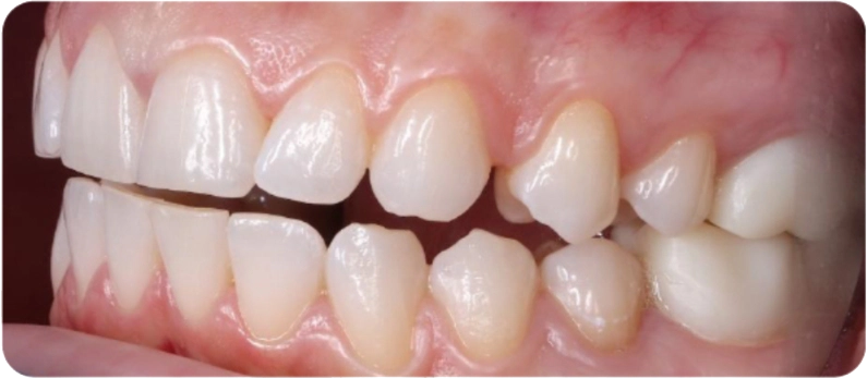
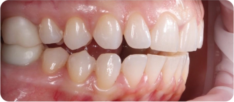
The patient presented with a distal occlusal relationship, reduced occlusal vertical dimension, unstable occlusion, and temporomandibular joint dysfunction.
Chief Concerns
The patient reported difficulty chewing, aesthetic concerns, and functional discomfort when bringing the upper and lower teeth into occlusion.
Treatment Objective
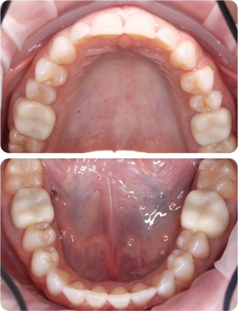
A comprehensive digital assessment was performed to establish an appropriate therapeutic mandibular position. Four ceramic crowns were then digitally designed and 3D printed to restore posterior contacts, improve occlusal stability, and support masticatory function.
Restoration Sites
Bilateral maxillary and mandibular first molars (4 crowns total)
Digital Workflow for Occlusal Rehabilitation
01 Data Acquisition and Diagnosis
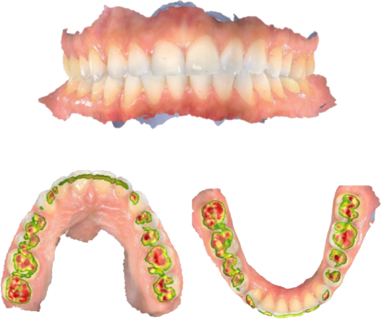
The first priority was to identify a mandibular position that could support a stable, functional bite. Prof. Apresyan combined intraoral scan data with CBCT imaging and a functional TMJ assessment to understand how the patient’s reduced vertical dimension and joint condition were affecting the occlusion.
Rather than reproducing the existing bite, the digital records were used to plan a new pattern of posterior support. Four first molars were selected for restoration. Their occlusal height and anatomy were redesigned to create more balanced posterior contacts at the planned mandibular position.
02 Digital Design
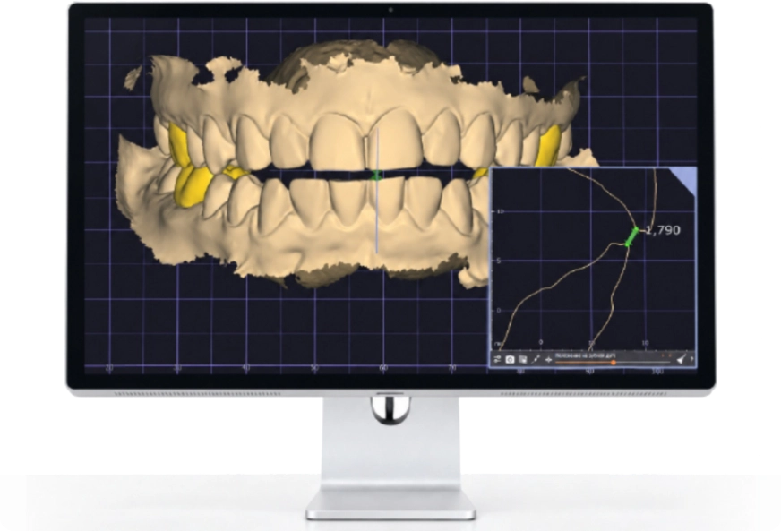
The four ceramic crowns were designed as a coordinated occlusal set rather than as isolated restorations. This allowed Prof. Apresyan to shape the posterior contacts around the planned mandibular position and vertical dimension.
During the design process, particular attention was paid to:
-
Margin definition and material thickness
-
Cusp and fossa anatomy
-
Contacts with the opposing dentition
-
Proximal contacts
-
The planned occlusal vertical dimension
03 3D Printing and Post-Processing
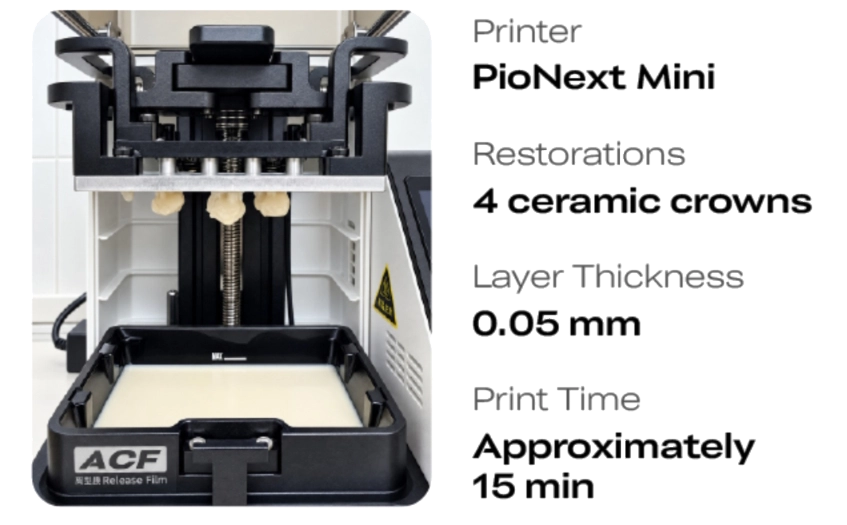
Once the designs were finalized, all four crowns were nested in a single build and printed on the PioNext Mini.
Printer: PioNext Mini Restorations: 4 ceramic crowns Layer Thickness: 0.05 mm Print Time: Approximately 15 minutes
The precise reproduction of crown morphology — particularly margins and occlusal anatomy — is critical for optimal passive fit and occlusal contact. For Prof. Apresyan, accuracy, stable performance, and a high print success rate are therefore essential when choosing a dental 3D printer.
In this case, the PioNext Mini successfully fabricated all four crowns in a single print run. The resulting restorations exhibited exceptional fidelity to the digital design, flawlessly reproducing intricate anatomical details. After simple post-processing, they were ready for clinical try-in.
“What impressed me most about the PioNext Mini was its accuracy and stable printing performance. The printed crowns reproduced the fine details of the designs and required only simple post-processing before try-in.”
— Prof. Samvel Apresyan
The PioNext Mini also features an intuitive interface and a simple workflow. Once the digital designs are ready, printing can begin quickly, making the system easy to use in everyday clinical practice.
Ready for Same-Day Try-In
In approximately two hours, Prof. Apresyan moved from digital data collection to four try-in-ready crowns, with printing completed in about 15 minutes. The crowns fit well and required minimal chairside adjustment.
By producing them in-house, the patient could begin bite adjustment on the same day without waiting for an external lab. The restored posterior support also provided a stable starting point for the next stage of treatment.